EKG 2

Here’s another EKG to get you guys talking. Thank you Dr. Silverberg and Dr. Brown for the case.

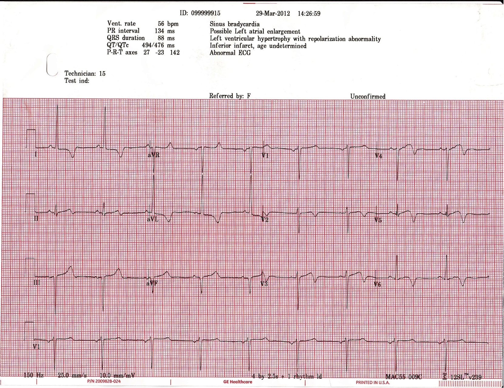
73 chinese female h/o dm, htn and high cholesterol had syncopal episode. Felt heart racing and palpitations prior to passing out at bingo in her church. Only complaint now is slight chest pain that is similar in nature to what she felt prior to today’s syncope only it was worse then. Vitals: hypertensive but the rest normal including the physical.


What’s the diagnosis?

What’s your management?


The views expressed on this blog are the author's own and do not reflect the views of their employer. Please read our full disclaimer here. Any references to clinical cases refer to patients treated at a virtual hospital, Janus General Hospital.
The following two tabs change content below.

jwillis

James Willis, MD. Assistant Program Director at SUNY Downstate / Kings County.

Latest posts by jwillis (see all)

4 comments for “EKG 2

  1. Richard.Shin
    April 5, 2012 at 10:48 am

    This EKG is concerning. The T waves in the anterior, lateral and inferior leads are inverted and some are biphasic. The ST segment is not concave. My suspicion for an acute MI is very high.

    I would give aspirin, give nitro for chest pain, cardiac labs, try and get an old EKG, repeat ekg with any episode of chest pain, early cardiology consult, pacer pads attached and would make sure they are being placed on continous cardiac monitoring.

    • Richard.Shin
      April 5, 2012 at 4:25 pm

      Just to add it looks like wellen’s warning or wellen’s syndrome which can be a sign of proximal LAD occlusion, would definately NOT stress this patient, would push for direct catheterization

  2. doty
    April 5, 2012 at 11:28 am

    Another great EKG. It should make you nervous. We can’t really call this a STEMI. however. the morphology of the ST changes is very concerning. Maybe we should stress him to see if has reversible CAD? Good idea?

    I am sure some of the studs out there will nail this case.

  3. mritchie
    April 6, 2012 at 10:32 am

    Read:
    Rhythm: Sinus
    Rate: 56
    Axis: Left axis deviation (-23)

    P-waves: inverted in V1 suspicious for LAE
    PR Interval: Normal (134)
    QRS: Narrow (88)
    Q waves: III and aVF, suspicious for old inferior MI
    R waves: Good progression V1-V6
    LVH: Meets Cornell’s Voltage critieria
    ST segment: No elevations or depressions
    T waves: Flipped in I,aVL,V2,V6
    Biphasic in V3-V5

    Answer: LAD coronary T-wave syndrome (Wellen’s Syndrome)

    First described in 1980’s by Wellen

    Criteria:
    Progressive symmetrical deep T wave inversion in leads V2 and V3 a variant is biphasic T waves
    Little or no cardiac marker elevation
    Discrete or no ST segment elevation
    No loss of precordial R waves.
    Pattern abnormal during chest-pain free periods

    As seen in eMedicine:
    “Wellens syndrome represents critical stenosis of the left anterior descending coronary artery (LAD). LAD’s nickname “widow maker.” The LAD supplies the anterior wall of the heart, including both ventricles, as well as the septum. An occlusion in this vessel can result in serious ventricular dysfunction
    Evolution to an anterior wall infarction is rapid, with a mean time of 8.5 days from onset of Wellens syndrome to infarction.”

    These patient’s usually have at least 50% stenosis if their LAD and should not undergo stress test. These patient should receive angiography for possible PCI or CABG.

    That’s what I came up with. Any other thoughts. I think Shin was correct.

Comments are closed.