Thanks to Dr. Caputo for today’s Morning report!
Hand Infections
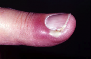
Recent trauma to the lateral nail fold
Acute: History of nail biting or manicuring, finger sucking
Chronic: Repeated exposure to water and/or irritants
Erythema and pain in early stages, followed by frank abscess formation with increased pain
History of diabetes
Felon
Recent trauma to finger pad
Pain, typically throbbing in nature
Swelling, pressure, and erythema
Presence and/or progression from paronychia
Herpetic whitlow
Genital herpes in self or partner
Health care worker , Children with gingivostomatitis
Most commonly on tip of finger rather than on shaft
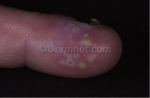
Localized pain, pruritus, and swelling followed by the appearance of clear vesicles
Typically localized to 1 finger only (involving > 1 finger are more typical of coxsackievirus infection)
Infectious tenosynovitis
Recent penetrating trauma to the hand
Gonococcal infection, particularly disseminated
Pain, especially with passive extension of the finger
Edema of the entire finger
Variable history of fever, Kanavel’s signs
Deep fascial space infection
Recent penetrating trauma to the hand
Recent or untreated tenosynovitis
Palmar blister (subfascial web space abscess)
Pain and edema of the hand
Pain with movement of the fingers
Variable history of fever
Physical Exam
Paronychia
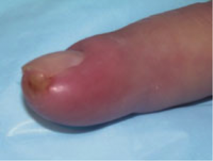
Edema, erythema, and pain along lateral edge of the nail fold
May have extension to the proximal nail edge (eponychium)
Presence of frank abscess formation and fluctuance, although less common in chronic cases
Subungual abscess (floating nail) if pus has extended under the nail plate
Felon
Painful, tense, and erythematous finger pad
Pointing of abscess possibly present
Signs typically limited to area distal to the distal interphalangeal joint because of anatomic constraints
Evidence of penetrating trauma
Clear vesicles on an erythematous border localized to one finger
Pain, typically out of proportion to findings
Edema
Turbid or cloudy fluid in vesicles, possibly suggesting a superimposed pyogenic infection
In later stages, coalescence of vesicles to form an ulcer
Distal finger pulp that remains soft, which may help distinguish herpes simplex virus (HSV) infections from bacterial felons
The 4 cardinal signs, first described by Kanavel, include the following:
Tenderness along the course of the flexor tendon
Symmetric edema of the involved finger
Pain on passive extension (believed by some authors to be the most important sign)
Flexed resting posture of finger
All 4 signs are possibly not present early in the course of infection. Patients may have associated lymphangitis, lymphadenopathy, and fever.
Deep fascial space infections all possibly present with lymphangitis (identified dorsally), lymphadenopathy, and fever.
Dorsal subaponeurotic abscesses – Result in swelling and pain on the dorsum of the hand and pain with passive movement of the extensor tendons (difficult to distinguish from dorsal subcutaneous infection)
Subfascial web space infections – Present with pain and swelling on the dorsum and palmar surfaces of the hand (Because the subfascial space is contiguous with the dorsal subcutaneous space, tracking of infection from the former to the latter results in a collar button abscess named for its hourglass shape.)
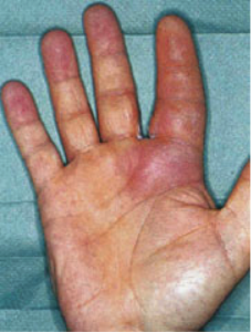
Midpalmar space infections – Present with pain, swelling, loss of palmar concavity, pain with movement of the third and fourth digits, and dorsal swelling secondary to the tracking of infection dorsally along the lymphatics
Thenar space infections – Result in marked swelling of the thumb-index web space, flexed and abducted resting posture of the thumb, and pain with passive adduction
Causes
Paronychia
Staphylococcus aureus and Streptococcus species are most common in acute cases.
Candida albicans (95%) and atypical mycobacteria are causes in chronic cases and in patients who are immunocompromised.
Anaerobes may be involved in the pediatric population secondary to finger sucking and children’s playing in unhygienic spaces.
Felon
S aureus is the most common causative organism, but gram-negative organisms have been identified.
Herpetic whitlow
HSV-1 or HSV-2 is responsible.
Infectious tenosynovitis
S aureus and Streptococcus species are commonly isolated; however, some authors believe that N gonorrhoeae should be considered a possible pathogen until excluded by culture data.
Eikenella corrodens is observed in infections caused by human bites.
Pasteurella multocida and Capnocytophaga infections caused by cat and dog bites can progress rapidly to septic shock and death.
Deep fascial space infections
S aureus and Streptococcus species are most commonly isolated.
Organisms mentioned for infectious tenosynovitis also apply to deep space infections. This may be the result of local spread from infected neighboring tendon sheaths.
Treatment
Paronychia
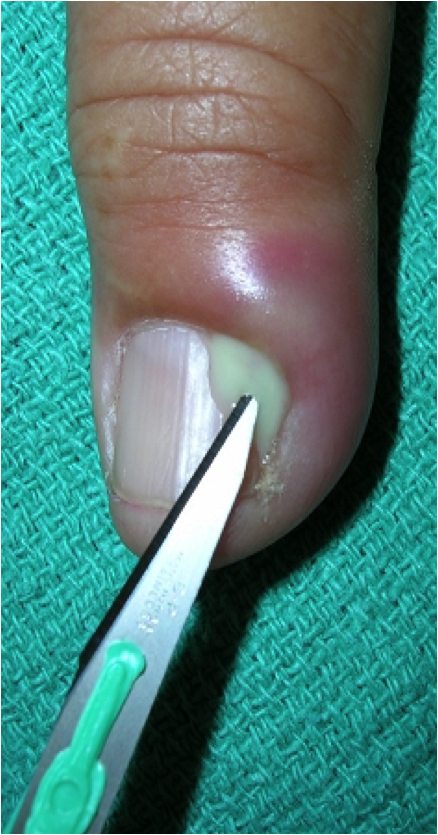 In acute paronychia, if no frank abscess or fluctuance is noted along the lateral nail edge, frequent hot soaks and possibly a short course of antibiotics may result in resolution of the infection.
If pus is present, drainage of the area is required. Perform a digital nerve block using lidocaine without epinephrine. Using a No 11 scalpel blade held parallel to the nail, elevate the lateral nail fold at the site of the abscess to allow for drainage of pus. If a large amount of pus is expelled, a small wick is left in the incision to allow for continued drainage. If pus has tracked beneath the nail, the removal of an adjacent longitudinal section of the nail may be necessary to promote drainage. If a subungual abscess results in a floating nail, remove a portion of the nail or trephinate the nail to allow for complete drainage.
After drainage and wick placement, dress the finger appropriately.
Update tetanus booster status as needed.
In chronic paronychia, treatment consists of avoiding predisposing factors and initiating topical steroids and antifungal agents. Surgical intervention is indicated only if medical treatment fails.
Felon
If frank abscess formation is present or the finger pad is tense, incision and drainage is indicated. Th
 is should not be undertaken lightly because improper placement of the incision can lead to scarring, sensory loss, unnecessary pain, instability of the finger pad, and spread of infection into the adjacent tendon sheath.
Elevation of the eponychial fold with a No 11 blade is quick, usually painless, and effective, and if there is pain it is extremely brief, less so than the pain of a digital block so local anesthesia is not typically needed. Discuss the procedure with the patient to alleviate their fears. Extensive incision or penetration of the finger with the blade is unnecessary, simple elevation of the fold will do; therefore, no nerve block is needed. If the patient requests it, a digital nerve block can be performed for comfort.
A longitudinal incision over the area of greatest fluctuance is the safest procedure when incision and drainage is required. Manyother procedures, including hockey-stick or fish-mouth shaped incisions, are no longer recommended because of injury to neurovascular structure.
To avoid penetration of the tendon sheath, the incision should not extend to the distal interphalangeal crease. Using a hemostat, bluntly dissect the wound to promote drainage. Irrigate the cavity copiously and loosely pack with a gauze wick. After irrigation and loose packing of the wound, apply a dry gauze dressing and overlying splint. Update tetanus booster status as needed.
Herpetic whitlow
Apply a dry gauze dressing to the affected finger to prevent further spread of the lesion.
Infectious tenosynovitis and/or deep fascial space infections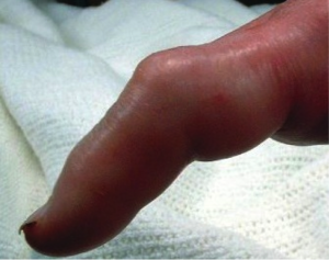
ED care consists of making the correct diagnosis, providing pain relief, initiating antibiotic therapy, elevating and immobilizing the hand, and consulting an experienced hand surgeon promptly for definitive treatment. Experienced surgeons in the operating room should perform the incision and drainage.
Jay Khadpe MD
Latest posts by Jay Khadpe MD (see all)
- Morning Report: 7/30/2015 - July 30, 2015
- Morning Report: 7/28/2015 - July 28, 2015
- IN THE STRETCHER INSTEAD OF BESIDE IT - July 22, 2015
- Morning Report: 7/14/2015 - July 14, 2015
- Morning Report: 7/10/2015 - July 10, 2015

