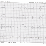
Here is the new EKG. There is a gift card prize to the person with the most complete answer. Look at Dr. Silverberg’s website for a guide on how to answer. Good luck.
The views expressed on this blog are the author's own and do not reflect the views of their employer. Please read our full disclaimer here. Any references to clinical cases refer to patients treated at a virtual hospital, Janus General Hospital.
The following two tabs change content below.


mritchie
Latest posts by mritchie (see all)
- X-ray Vision: The answer - July 31, 2013
- X-ray Vision: Ortho - July 17, 2013
- X-ray Vision Answer: - April 17, 2013
- X-ray Vision: Stories in the chest x-ray - April 2, 2013
- Rhythm Nation: Case 6 Answer - March 23, 2013


Rate: 60
Rhythm: NSR
Axis: left axis deviation
Intervals PR= approx 240ms (>200ms)=Prolonged PR, QRS >120ms
Morphology: RBBB QRS>120 with RR1 in V1, LAHB= slurred S in Inferior leads; RBBB+LAHB+1st Degree AV block= Trifascicular Block
ST segments: No elevations or depression
T Waves: Not flipped or flattened.
no history? i would look/ask for empty pill bottles…and have some bicarb near by.
Looks like TCA overdose, as I’m sure Dave was getting to.
There is a widened QRS, terminal R in AVR, and a deep S wave in I. All of which are classically seen in TCA overdose.
I think that is a great thought. It is always important to look at the whole EKG. AVR is often overlooked. While a terminal 40 R in AVR is seen in TCA overdose it is also seen in other diseases as well. RBBB is one of those. Below is the formal definition of RBBB.
Right Bundle Branch Block
RSR’ in V1-2
Broad S in I or V6
Broad R in aVR
TWI in V1 or V2; sometimes ST depression there too
Keep coming with guesses and as always, credit will only be given for formal guesses in the appropriate format.
Someone BETTER have gotten this!!! (see ED-ICU conference last month.)