
So we have an 81 yo W found apneic and cyanotic and EMS was called and patient was intubated. Patient had never been here before and no history came with the patient. The history was obtained with this chest x-ray.
The x-ray was done:
I think Grock did a good job of a formal read of this chest x-ray. It is a good idea to approach x-rays in this fashion so you don’t end up with errors.
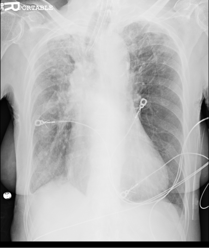
It is an AP film; portable
I will use Andy’s format but put in the official answer.
A; Airway: Diffuse circumferential pleural parenchymal disease throughout the right lung with volume loss causing superior retraction of the right hilum. Apical scarring and fibrosis
B; Bones: posterior left rib fractures
C; Cardiac: enlarged cardiomediastinal silhouette
D; Diaphragm: small right pleural effusion
When looking at the chest x-ray we came to the conclusion of interstitial lung disease and probable history of Tb due to RUL scarring, It was later found that her only medical problems were ILD and has a history of Tb that was treated.
Bottom line: Chest x-rays can hold a lot of information.
So congratulations to Andy Grock for the best, most complete answer.
mritchie
Latest posts by mritchie (see all)
- X-ray Vision: The answer - July 31, 2013
- X-ray Vision: Ortho - July 17, 2013
- X-ray Vision Answer: - April 17, 2013
- X-ray Vision: Stories in the chest x-ray - April 2, 2013
- Rhythm Nation: Case 6 Answer - March 23, 2013