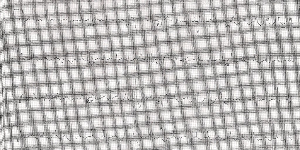
 Here is the answer for our last EKG:
Rate: 300
Rhythm: Sinus rhythm – although the P waves are difficult to discern in the rhythm strip, they are visible in V1. Mapping the P waves in V1 to the rhythm strip, you can see that the P waves in lead II are downgoing waves that occur immediately after the end of the T wave. The wide complex beats that occur at irregular intervals are PVCs.
Axis: Normal axis
Intervals: PR about 160ms; QRS borderline at 120ms with a RBBB pattern; QT about 280ms which gives QTC about 430ms
ST segments: T waves are discordant with the QRS in almost every lead. No ST segment depressions or elevations
Chambers – no signs of LVH or atrial enlargement; The negative P wave in inferior leads may indicate an ectopic atrial pacemaker causing abnormal direction of conduction of the atrial impulse.
This is a case of atrial tachycardia. The P wave is originating from an ectopic focus. The atrial rate is fast (>100bpm). The paves are consistent throughout the EKG which makes multifocal atrial tachycardia unlikely. The baseline is isoelectric, which makes atrial flutter less likely.
The treatment of atrial tachycardia varies depending on the cause and the stability of the patient. Unstable patients may require cardioversion. Antiarrhythmic therapy is also an option, and catheter ablation may be considered in patients with recurrent episodes and a single ectopic focus.
Thanks to Carl for submitting this EKG!
nchristopher
Latest posts by nchristopher (see all)
- What’s wrong with this picture? – Answer - September 11, 2013
- What’s wrong with this picture? - August 21, 2013
- EKG Case 8 – Answer - July 16, 2013
- EKG Case 8 – All that wheezes - June 19, 2013
- EKG Case 7 Answer - June 19, 2013

Nice ECG and review. Some more:
The key is the consistent AND abnormal p wave morphology. If you look at lead II the p wave is primarily negative (downward) in its vector and a normal p would be positive (upward). This means there is an abnormal p axis which implies an ectopic atrial focus.
The mechanism of an ectopic atrial tachycardia is abnormal automaticity – that is, a group of cells generate rhythmic impulses when they are not supposed to. Automatic dysrhythmias WILL NOT respond to cardioversion. Increased sympathetic tone often triggers these rhythms, so beta-blockers is a good first choice of medications. Definitive treatment involves catheter ablation.