EKG Case 8 – All that wheezes

ekg
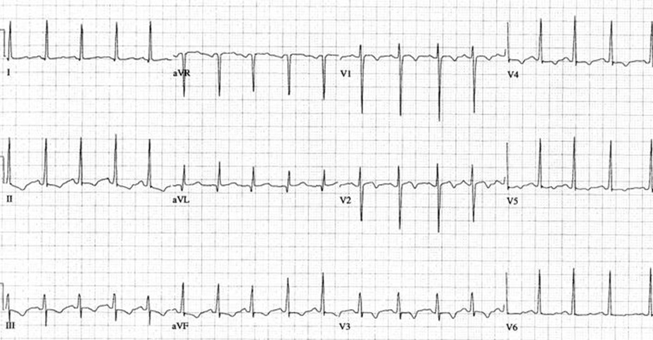
24yo F presents with SOB, wheezing and chest tightness.  Exam shows tachycardia and wheezes.  The above EKG is obtained.  What is your DDx?  Any other information about the patient you would like to know?


The views expressed on this blog are the author's own and do not reflect the views of their employer. Please read our full disclaimer here. Any references to clinical cases refer to patients treated at a virtual hospital, Janus General Hospital.
The following two tabs change content below.

nchristopher

Latest posts by nchristopher (see all)

2 comments for “EKG Case 8 – All that wheezes

  1. andygrock
    June 26, 2013 at 11:45 pm

    I’m would be more offended that more people haven’t responded, but hey, I guess the gift card is mine.

    DDx from history/physical: pericarditis (my favorite is Tb pericarditis), myocarditis cor pulmonale, eisenmengers/chf, asthma, PE, pulmonary htn (hereditary vs from OSA), pna, crack lung, COPD (likely from alpha 1 trypsin insufficiency), ACS (higher risk due to sickle cell anemia/family history), restrictive lung disease due to severe scoliosis, cardiac contusion s/p trauma, atelectasis leading to V/Q mismatch from…chronic heroin use/rib fx/being fat.

    EKG: Rate: 12ish
    Rhythm: regular, sinus
    Axis: nml axis
    Intervals: QRS nml (if you squint hard and make believe maybe a delta wave seen best in AVF), PR short to nml (hard to see little lines), QT likely nml (looks nml in AVL, but difficult to tell where T wave ends in II/III making QT calculation difficult)
    T waves: inverted in II/III/AVF, V1/V2/V3/V4
    P waves: possibly different waveforms…(MAT?) P wave in II appears abnormal, but is upright).
    ST-T – no obvious ST elevation/depression.
    No electrical alternans
    Decent R wave progression

    General interpretation: sinus tach (pain/PE/CHF?), MAT, Very soft call for wpw,

    Things I want: Prev EKG, PMHx, is she fat?, fever?, recent travel?, PQRST for symptoms?, tachypnea?, PERC/Wells score, Echo done by Dr. Christopher (I hear he gets apical 5 every time…), CXR, ABG/O2 sat.

  2. mritchie
    July 2, 2013 at 5:48 am

    My interpretation:
    Rate: 114
    Rhythm: Sinus Tachycardia
    Axis: Normal

    P waves: no signs of atrial enlargement
    PR interval: normal
    QRS: Narrow
    No Q waves
    LVH by criteria
    Good R wave progression
    ST segments: no elevation or depression
    T waves: Flipped in II, III, AVF, V1,V2,V3,V4,V5

    My guess on the diagnosis would be pericarditis.

Comments are closed.