Brought to you by Dr Andrew Grock and Dr. Elizabeth Abram. Supervised by Dr Martindale.
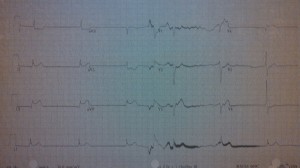
A nurse brings you the following EKG to sign. You go to the patient’s bedside:
63 yo F, pmhx dm/htn, here for epigastric pain, vomiting, dizziness x 4 hours. She is diaphoretic and reporting active pain and nausea at this time.
To win the week’s prize please provide the following:
1. Full interpretation of ekg including rate, rhythm, axis, intervals, st-t segments, t- wave interpretation, other
2. Please explain pathophysiology of the ekg abnormality or abnormalities featured above
3. Please provide the appropriate sequence of treatment actions in caring for this patient.
4. Which common treatment for this problem is contra-indicated?
The answer will be posted next Friday!
andygrock
- Resident Editor In Chief of blog.clinicalmonster.com.
- Co-Founder and Co-Director of the ALiEM AIR Executive Board - Check it out here: http://www.aliem.com/aliem-approved-instructional-resources-air-series/
- Resident at Kings County Hospital
Latest posts by andygrock (see all)
- A Tox Mystery…. - May 26, 2015
- Of Course, US Only for Kidney Stones… - May 18, 2015
- Case of the Month 11: Answer - May 12, 2015
- Too Classic a Question to Be Bored Review - May 5, 2015
- Case of the Month 11: Presentation - May 1, 2015

Looks like inferior MI, most likely due to RCA lesion (circumflex lesions usually will have higher STE in II than III). I can’t see any P waves, I’m not sure if that’s because of the quality of the image. She is markedly bradycardic as well, in conjunction with the inferior MI she probably has a complete heart block due to ischemia of the conduction system. If she is hypotensive I would give her fluids and then place a temporary pacer, while activating the cath lab. Nitrates are contraindicated.
The bradycardia may also be mediated by the bezold jarisch reflex
Looks like big inferior MI, but I think possibly LAD lesion as well with STE in aVr, The precordial leads are tough to see. The reciprocal changes fit with just inferior MI. I would just say big RCA lesion. This looks no no atrial rhythm at all to me. I think its a ventricular escape rhythm. This is badness. I’m sure this is peri-arrest.
these are big lesions caused by an acute mi
agree with nathan on management. Definitely no nitro. Emergent cath or lytics if you’re going to have to transport. I would also use small fluid bolus to volume expand as much as I can while waiting watching for fluid overload in the lungs. Maybe balloon pump if they have no EF, but that’s for later as well. In the ER waiting for cath you may need pressors.
We better be ultrasounding this guy. I want to know ef, lung and maybe ivc.
You can see similar pattern with LAD but my money is on RCA because of how high III is, with left sided lessons lead II will typically have greater STE than III.
Look at the big brains on these ^^ guys….. Nice.
If suspecting MI, then many would discourage atropine as well.
According to guidelines, rate can be treated with EITHER pacing OR inotropy until definitive intervention of TPA or PCI.