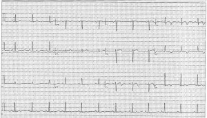
45 yo nulliparous F of Hispanic and Caucasian descent pmhx HTN on longstanding ARB p/w left intermittent arm pain x 1 year and 2 days of worsening mid back discomfort and SOB. Pt regularly walks 5 miles a day and has a BMI of 22. Had a benign bladder polyp resected 3 months prior and since then has entered perimenopause with irregular menses. Denies nausea/vomiting/diaphoresis. No other medical problems or environmental exposures.
SH: occasional ETOH (1-2 glasses of wine/wk), no smoking or illicits
FH: one aunt deceased prior to age 50, no known CAD
VS: 152/98, HR 72, RR 16, SaO2 100%, afebrile
PE otherwise unremarkable… except you note the patient seems rather flexible for her age.
EKG as above.
CXR unremarkable.
This patient was sent from her PMD via ambulance for the above abnormal EKG.
- What about this EKG is concerning?
- After a troponin of 1, Cardiology does a bedside echo and discovers wall-motion abnormalities. The patient is taken to cardiac cath. No plaques are seen, yet stents are placed. What is the location(s) of the stents?
- What is this condition called?
- What are some of the underlying conditions that may have pre-disposed this patient to her diagnosis?
Now is your chance to figure out what was going on! Best answer to the above 4 questions by noon Friday 7/18/14 wins.
By Dr. Elizabeth Abram
Supervised by Dr. Jennifer Martindale.
eabram
Latest posts by eabram (see all)
- Rhythm Nation May 2015 Answer! - June 1, 2015
- Rhythm Nation May 2015 - May 10, 2015
- Rhythm Nation April 2015 – Answer! - April 20, 2015
- Rhythm Nation April 2015 - April 13, 2015
- Rhythm Nation March 2015 – Answer! - March 31, 2015


This could be the arterial form of EDS with coronary artery aneurysm. The ECG itself makes me think of PE but the clinical scenario makes me more suspicious for EDS, especially since you mention that a stent was placed but there were no plaques.
Or coronary artery dissection
Cant say Ive ever ventured into the world of county blogging, but I didnt have anything particularly exciting to do today.
The EKG has biphasic Ts in anterior leads makes me think Wellens Syndroms (prox LAD occlusion) and would explain her anginal symptoms throughout the year. However, they found no plaque on cardiac cath which makes this answer wrong.
If you want to get really crazy you can consider Takotsubo (had to check spelling on that) which would cause acute heart failure with wall motion abnormalities and could present on EKG as an anterior wall MI (T wave inversions) with clean coronaries. Probably also wrong, but hey, I’m participating.
Nathan’s probably right with Ehler Danlos/coronary aneurysm given her flexibility but maybe she just does yoga?
1) This EKG is concerning because it points to an anatomic distribution of abnoramalities. Specifically there are biphasic T waves in the precordial leads (V3) and a deep T wave inversion in V4. As other commenters have pointed out this like a Wellen’s Sign
2) The abnoramility is therefore likely in the proximal LAD, similar to the lesion classically found in wellen’s syndrome
3) Spontaneous Coronary Artery Dissection
4) Hypertension. Female Gender. Hormones (did somebody give her OCPs or other exogenous hormones for her irregular menses?). Connective tissue disorder “rather flexible for her age” – could be a Ehler’s Danlos or similar varriant.