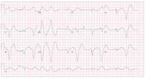
47 year-old female with past medical history of depression and supraventricular tachycardia, brought in by EMS with altered mental status and found with empty pill bottles nearby.
What is the interpretation of this ECG?
What likely happened to this patient?
How do you treat it?
Best answer by December 19, 2014 at noon will win!
The views expressed on this blog are the author's own and do not reflect the views of their employer. Please read our full disclaimer here. Any references to clinical cases refer to patients treated at a virtual hospital, Janus General Hospital.
The following two tabs change content below.


eabram
Latest posts by eabram (see all)
- Rhythm Nation May 2015 Answer! - June 1, 2015
- Rhythm Nation May 2015 - May 10, 2015
- Rhythm Nation April 2015 – Answer! - April 20, 2015
- Rhythm Nation April 2015 - April 13, 2015
- Rhythm Nation March 2015 – Answer! - March 31, 2015


What is the interpretation of this ECG?
Looks like a bad TCA overdose. EKG showing widdening QRS complexes to the point of near syne waves. Of note it’s not a regular rhythm, on the rhythm strip there are 3-4 beats of one morphology, followed by a more sinus rhythm for 3 beats. I think that is going on is the pt’s history of SVT, so likely the inital beats are re-entry via an accessory pathway.
What likely happened to this patient?
Took a blottle of Trycyclic Antidepressants.
How do you treat it?
Management would be for TCA tox:
–IV/O2/montior
–Sodium bicarbonate 100 mEq, repeat minutes until BP improves and QRS complexes begin to narrow.
–Intubate (airway protection)
–Activated charcol
–BP control with fluids, pressors if need be
–If arrhythmias occur, the first step is to give more sodium bicarbonate. Lidocaine (1.5mg/kg) IV is a third-line agent (after bicarbonate and hyperventilation) once pH is > 7.5.
Dispo: ICU
Great comment above. To add to it, TCAs are also anti-cholinergic which may also help explain the tachycardia seen in the ECG. They tend to increase the QT interval by potassium channel blockade. The reason lidocaine (Class 1b) is a good third-line agent is because of its effects on the action potential and how that affects the electrophysiology that is the basis of an ECG. (a nice picture on the right side bar depicts the changes described below http://www.cvpharmacology.com/antiarrhy/sodium-blockers.htm)
Class 1a antiarrhythmics cause prolongation of the QRS and QT interval by their action on Phases 0 and 3 of the ventricular action potential. Class 1c antiarrhythmics cause prolongation of the QRS interval by their action on the Phase 0 action potential. This is bad because, as is shown by the above example, TCA overdose prolongs duration of both these intervals.
Class 1b antiarrhythmics (e.g., lidocaine) decrease the QT by their action on shortening Phase 2 of the ventricular action potential leading to some correction of the ECG changes (cardiac electrophysiology).
In addition, TCAs may lead to seizure activity, which means the patient should also be monitored for such a development. Appropriate treatment consists of benzodiazepine therapy.
I like TCAs/Na Channel Blocker in the DDx. But there are some findings that don’t fit.
First off, this is not a classic TCA ECG. (see http://lifeinthefastlane.com/ecg-library/basics/tca-overdose/). This patient isn’t tachycardic, the axis isn’t rightwards, there’s no big obvious pathognomonic terminal R in AVR….
Let’s go systematically: This is a wide, irregular rhythm, with a rate in the 60s. PArts appear regular, parts are regularly-irregular. There are two different wide complex morphologies. There are no easily discernible p waves. The differing morphologies complicate the axis, which appear left-ward. Beats 1,3,4,5 are the same, are incredibly-wide. In the last half, the wide-beats appear to be coupled.
I think Grundmann is right to use the term “Sine”-wavy. This super-wide morphology begs consideration of toxic and metabolic causes. The sine-wave makes me think hyper-K.
The coupled wide-complexes without obvious abberancy or p waves makes me think these are two-different ventricular foci consistent with bidirectional Vtach. We’ve just learned from recent M&Ms that Bidirectional V tach has other causes than dig toxicitiy, but this is nearly pathognomonic.
The slow rate and the history of SVT fits with dig toxicity. Dig toxicity causes hyperK, so the sine-wave fits too.
Treatment: ABCs, digi-fab, +/- lidocaine, phenytoin, magnesium. +/- Atropine, cardiac pacing, chronotropes. ICU,