Case Recap: Old lady with hypotension, tachycardia and JVD…
I think we all agree that this person is sick. Prior to pondering the wide differential do the Basics: 2 Large Bore IVs, O2 NC vs NRB, Monitor, assess airway
My Top Three Differential Diagnoses
1. Cardiac Tamponade
2. Massive PE
3. Inferior Wall MI with Cardiogenic Shock
Do you want to give fluids?
Tricky question! IV fluids helps BP in PE and Tamponade…but can be a poor idea in cardiogenic shock. Remember, in cardiogenic shock the heart can’t pump the fluid out leading to decreased venous return as demonstrated by JVD. Fluids in cardiogenic shock are generally a bad idea as they increase an already overloaded pre-load. Since her lungs are clear iIn this case, you can give a “100ml to 250ml bolus” [1] and reassess the patient for improved BP and the development of rales.
Once Basics Are Done
ECG and Echo. Yes, labs/cxr are also needed, but they won’t help you differentiate between the above three in a timely fashion.
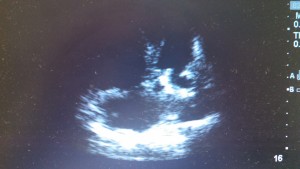
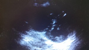
Her Echo may have looked like this…
Apical 4Parasternal Short
Parasternal Long
What findings are on these US images?
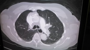
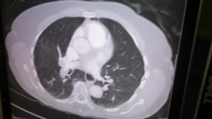
If the patient improves enough to get a CT...
Treatment?
By Dr. Andrew Grock
Special thanks to Dr. Ian deSouza.
References
[1] Tintinalli’s 7th ed.
andygrock
- Resident Editor In Chief of blog.clinicalmonster.com.
- Co-Founder and Co-Director of the ALiEM AIR Executive Board - Check it out here: http://www.aliem.com/aliem-approved-instructional-resources-air-series/
- Resident at Kings County Hospital
Latest posts by andygrock (see all)
- A Tox Mystery…. - May 26, 2015
- Of Course, US Only for Kidney Stones… - May 18, 2015
- Case of the Month 11: Answer - May 12, 2015
- Too Classic a Question to Be Bored Review - May 5, 2015
- Case of the Month 11: Presentation - May 1, 2015


Careful with IV fluid and consider early use of norepinephrine.
“Although patients with RV failure are often preload dependent, volume loading has the potential to overdistend the ventricles and cause increased wall tension, decreased contractility, increased ventricular interdependence, impaired LV filling, and reduced systemic cardiac output.”
Piazza G and Goldhaber SZ. The acutely decompensated right ventricle. Chest 2005; 128: 1836-52.
desouza, Oren Friedman was talking about limiting fluids this week at the critical care conference. However I’ve had hypotensive PE pts turn around quickly with 1 or 2 L of fluid.
Hey, couple points bout that echo.
The RV is huge and very thin walled.
The thickness of the RV wall can help you determine acute vs chronic elevated pulmonary pressures. You’ll get hypertrophy with the R wall with elevated pulmonary pressures just like you get a big LV wall from systemic hypertension. This is really big pressures and really acute.
The Left Ventricular Ejection Fraction looks good and no real L sided wall abnormalities, which goes against Myocardial Infarction (not that you’re really scratching your head for this one, but this helps in equivocal cases). You can see the LV starting to collapse in late systole.
With POC ultrasound, you should assess the lungs and IVC too while you’re looking at an undifferentiated dyspneic or hypotensive patients. Look for signs of fluid overload from a backed up LV and at the IVC to check for volume status (really only good in extremes of collapsing vs plethoric). I’m sure this lady’s IVC was huge.
anyway, great case.