Rhythm Nation January 2015

Happy New Year to All!

Now, back to business…

A 71 year-old male with past medical history of CAD and CHF (last known EF 10%) is sent by his PMD for progressive shortness of breath, dyspnea on exertion, and lower extremity edema.

Just as you sit down to finally catch up on notes, you are handed the ECG below.

January 2015

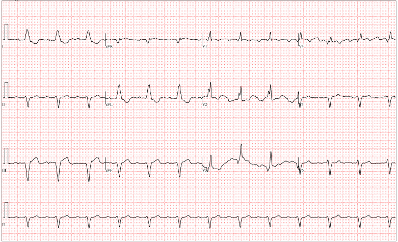
What is your interpretation of this ECG?

What type of conduction abnormality would you be most concerned for and why?

Briefly, how would you manage this patient in the Emergency Department?

Best response by 12pm on 1/16/15 wins!


The views expressed on this blog are the author's own and do not reflect the views of their employer. Please read our full disclaimer here. Any references to clinical cases refer to patients treated at a virtual hospital, Janus General Hospital.
The following two tabs change content below.

Yonatan

Latest posts by Yonatan (see all)

2 comments for “Rhythm Nation January 2015

  1. Nicolas Grundmann
    January 10, 2015 at 6:47 pm

    Rhythm: Regular Sinus
    Axis: Left axis deviation around –60 (upright in I, downward in AVL, iso in AVR)
    Rate: 75 bpm

    Blocks:
    1) 1st degree AV block with consistently prolonged P-R interval.
    2) Anterior hemiblock? No Q in I but a very deep S in III
    3) RBBB (widened QRS, R,R’ in V2)

    So this would be a Tri-facicular block, possibly from a occluded anterior descending branch of the LCA

    Management would be
    –IV / O2 / cardiac Monitor
    –ASA
    –cardiac enzymes / CBC / Chem-8 / type screen / coags
    –STAT cards consult vs STEMI code depending on the prior EKG (if this is new compared with prior EKG then call it as a STEMI code)
    – Have pacer pads there ready just in case

  2. boiyemhonlan
    January 14, 2015 at 1:47 am

    I agree with Nico’s read of the EKG. Dx: Trifascicular Block
    Rate: 75 bpm; Rythm: NSR; Axis: Slightly more neg aVL (-30 and 60)
    PR: Prolonged > 200 msec
    QRS: Wide rSR’ in V1 and V2
    ST: Normal, T wave (non specific inversions); QT normal
    First Degree Block + RBBB + LAD (Left Anterior Fascicle Blocked) = Trifascicular Block

    Patterns of Trifascicular Blocks include: Incomplete and Complete

    Incomplete – Fixed block of two fascicles w/ evidence of delayed conduction in the remaining fasicle (i.e AV block) or Fixed block of one fascicle w/ intermittent failure of two other fascicles (Bifas block + 1st degree block or Bifas + 2nd degree block, or RBBB + alternating LAFB/LPFB

    Complete block – Produces 3rd degree AV block with features of bifascicular block (escape rhythm arises from either the anterior or posterior fascicles producing QRS complexes with the appearance of RBBB + either LAFB or LPFB)

    I can’t determine which type of block the pt has but both are susceptible complete heart block. Nico’s management is spot on. May consider atropine, transcutaneous, or transvenous pacing based on vitals and clinical status. Obviously r/o other causes of heart block.

    (See LITFL for more information on this well as Dr. Martindale’s EKG Book.)

Comments are closed.