By Dr. Michelle DiMare
These days, the Pediatric ED is frequently filled with the cutest, most snot filled kids you’ve ever seen. With most of them reporting some degree of cough and fever, pneumonia is frequently considered on our differential diagnosis. While the incidence of CAP in developed countries is anywhere from 10 – 40 out of every 1000 children under 5, the majority of these patients don’t have pneumonia. But how can we tell the difference? More importantly, how do we decide who to treat with antibiotics? Should we use X-ray? Clinical suspicion? Treat ‘em all? Wait for them to be really sick?
As always, the answer is…ULTRASOUND!!
Recent guidelines suggest that in mild to moderate CAP, ie, cases that we would likely send home, clinical suspicion is enough to treat patients with antibiotics for presumed pneumonia. These patients don’t require any radiological tests for confirmation and rarely return with complications. We assume we correctly identified the patient’s pneumonia, treated appropriately and the patient got better. Chances are, however, that a large percentage of the patients would have gotten better even without our sharp clinical suspicion and antibiotics. A large portion of them probably have, at most, viral pneumonia, if not a simple URI.
Ultrasound is the perfect tool to help limit unnecessary antibiotics in children with suspected mild to moderate CAP.
Who should you ultrasound?
- anyone who you previously would have considered empirically treating
- ie persistent fever, isolated crackle, cough without other URI sx
- anyone who you would have gotten an xray on
- ie, unexplained tachypnea, cough for greater than 7 days
How should you ultrasound?
- use the linear probe
- scan in the sagittal plane
- scan the posterior lungs first, then anterior
- cover each intercostal space during at least 1 inspiratory/expiratory phase
What are you looking for?
Isolated B lines
- not diffuse or gravity dependent as in adults with CHF
- often only in 1-2 intercostal spaces
- a sign of interstitial or alveolar fluid
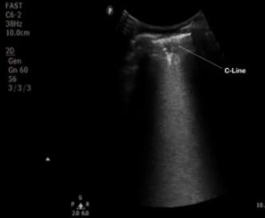
C lines
- a hypoechoic irregularity seen below the plural line
- arise from within consolidated lung tissue
Shred sign
- similar to C lines
- irregular line distinct from the pleural line arising from visceral pleura tethering
- large areas of “shred” at the base of the lung can be confused for a small pleural effusion
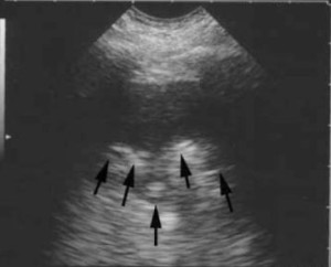
Air bronchograms
- linear branches created by air filled bronchi in otherwise consolidated, hypoechoic lung
- the air filled bronchi demonstrate bright, posterior acoustic enhancement making them appear hyperechoic
- are indicative of either pneumonia OR atelectasis
- dynamic à pneumonia
- static à atelectasis
- really difficult to determine dynamic vs static
- easier to use clinical context ie fever, cough to determine etiology
How to apply findings?
- any child with any of the above findings and even low suspicion of CAP should be treated
- any child with a normal lung ultrasound and good follow up can be observed as opposed to empiric treatment
Where’s the evidence?
- the evidence is somewhat limited because lots of studies compare US to CXR
- CXR is not the gold standard for diagnosing pneumonia
- No one is going to CT scan kids to find mild to moderate CAP
- US vs CXR with CXR as gold standard – good correlation
- Italian Journal of Pediatrics April 2014
- 48 of 103 with radiographically confirmed PNA
- Sens 97.9%
- Spec 94.5%
- PPV 94%
- NPV 98.1%
- US vs CXR with clinical assessment/British Thoracic Guidelines as gold standard
- Pediatric Pulmonology March 2013
- 89 of 102 found to have CAP by gold standard
- CXR 81/89
- US 88/89 –winner!!
- Pediatric Pulmonology March 2013
- Italian Journal of Pediatrics April 2014
How will this change your practice?
- decrease number of x-rays we’re getting on kids
- increase threshold for watchful waiting in children with fever and cough
- decrease antibiotic use in stable children without US findings of pneumonia
Thanks for reading and remember…if you have a fever, the only prescription is more ultrasound!!!!
References
Esposito S, Cohen R, Domingo JD, Pecurariu OF, Greenberg D, Heininger U, Knuf M, Lutsar I, Principi N, Rodrigues F, Sharland M, Spoulou V, Syrogiannopoulos GA, Usonis V, Vergison A, Schaad UB. Antibiotic therapy for pediatric community-acquired pneumonia: do we know when, what and for how long to treat? Pediatr Infect Dis J. 2012;31:e78–e85. doi: 10.1097/INF.0b013e318255dc5b.
Lichtenstein D, Meziere G. Relevance of lung ultrasound in the
diagnosis of acute respiratory failure, the BLUE protocol. Chest
2008; 134: 117-25
Caiulo, V. A., Gargani, L., Caiulo, S., Fisicaro, A., Moramarco, F., Latini, G., Picano, E. and Mele, G. (2013), Lung ultrasound characteristics of community-acquired pneumonia in hospitalized children. Pediatr. Pulmonol., 48: 280–287. doi: 10.1002/ppul.22585
Esposito S, Papa SS, Borzani I, et al. Performance of lung ultrasonography in children with community-acquired pneumonia. Italian Journal of Pediatrics 2014;40:37. doi:10.1186/1824-7288-40-37.
andygrock
- Resident Editor In Chief of blog.clinicalmonster.com.
- Co-Founder and Co-Director of the ALiEM AIR Executive Board - Check it out here: http://www.aliem.com/aliem-approved-instructional-resources-air-series/
- Resident at Kings County Hospital
Latest posts by andygrock (see all)
- A Tox Mystery…. - May 26, 2015
- Of Course, US Only for Kidney Stones… - May 18, 2015
- Case of the Month 11: Answer - May 12, 2015
- Too Classic a Question to Be Bored Review - May 5, 2015
- Case of the Month 11: Presentation - May 1, 2015


