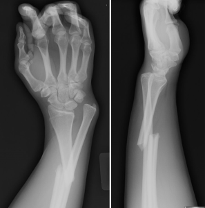
Here’s the image again
The answer is….Galeazzi fracture! Congrats to Dr. Berkowitz for the correct answer and treatment!
Galeazzi Fracture
Basics:
Galeazzi fractures are isolated fractures of the junction of the distal third and middle third of the radius with associated subluxation or dislocation of the distal radio-ulnar joint (DRUJ).
Mechanism is usually axial load on a hyperpronated forearm.
Presentation:
Patients typically presents with pain and soft tissue swelling at the distal radius and at the wrist.
Anterior interosseous nerve (AIN) palsy may be present. AIN is a purely motor nerve and is a division of the median nerve. If AIN is injured, paralysis of the flexor pollicis longus and flexor digitorum profundus will be noted at the index finger. At presentation, patient will be unable to pinch together the index finger and thumb.
Workup:
The fracture will be confirmed on plain radiograph. Both AP and lateral views of the forearm will need to be obtained.
Treatment:
All adult Galeazzi fractures MUST be treated with open reduction and internal fixation (ORIF). The fracture is termed “fracture of necessity” because its necessary surgical treatment. In adults, nonsurgical treatment will result in persistent dislocation of the distal ulna.
In children, these fractures can be treated with closed reduction and casting because of the skeletal immaturity of these patients.
Take Home Points:
Analgesia. Xray. Ortho consult for surgical management in adults and casting in children.
References:
http://emedicine.medscape.com/article/1239331-overview#a03
Tintinalli’s Emergency Medicine: A Comprehensive Study Guide
jwang
Latest posts by jwang (see all)
- Xray Vision Answer - May 24, 2015
- Xray Vision: My Arm Looks Funny…. - May 16, 2015
- Xray Vision: Limping Answer - April 27, 2015
- Xray Vision: Limping - April 17, 2015
- Xray Vision: Answer - March 27, 2015

