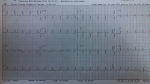
A 58 year old woman with a long medical history – paroxysmal afib, not on anticoagulation, hyperthyroidism secondary to graves disease, and GERD who presents for the third ED visit in 1 month for epigastric pain. At her first visit, she improved after a GI cocktail, had a normal ECG/CXR/labs, and was discharged with next day stress test. She returned with the same complaint, improved with another GI cocktail, had a normal ECG and CXR, and was discharged.
You see her on her third visit with the same presentation. Her vital signs are as follows: HR 120, RR 18, BP 110/61, O2 sat 99%, Temp 98.3 oral.
She is complaining of the same epigastric pain radiating to the chest with mild nausea that she has had for months. Additionally, she has been taking 8-10 ibuprofen tabs a day. Her ECG is…
After a GI cocktail and normal labs including a negative troponin, you go back to reassess the patient. She is now complaining of L chest pain and appears mildly diaphoretic. You put her on the monitor and now her HR is in the 150s.
For this month’s big prize
1. Please list your top 3-5 diagnosis, the next diagnostic step or steps, and the ED management for ?
andygrock
- Resident Editor In Chief of blog.clinicalmonster.com.
- Co-Founder and Co-Director of the ALiEM AIR Executive Board - Check it out here: http://www.aliem.com/aliem-approved-instructional-resources-air-series/
- Resident at Kings County Hospital
Latest posts by andygrock (see all)
- A Tox Mystery…. - May 26, 2015
- Of Course, US Only for Kidney Stones… - May 18, 2015
- Case of the Month 11: Answer - May 12, 2015
- Too Classic a Question to Be Bored Review - May 5, 2015
- Case of the Month 11: Presentation - May 1, 2015


58 year old woman with paroxysmal a fib, Graves disease, GERD has recurrent visits for epigastric pain now has chest pain, diaphoresis, and an EKG with diffuse ST elevations with PR depressions and sinus tachycardia. Her heart rate is 150 and although her paroxysmal a-fib might explain this in other circumstances, her rhythm is sinus. Volume depleted states rarely make someone this tachycardic, the fast rate is more likely to be primary cardiac or toxic/metabolic. Her labs were normal so metabolic derangements are less likely.
Differential:
#1: Pericarditis. The EKG is consistent with pericarditis, which is also supported by the symptoms of chest pain. Pericarditis is associated with Graves disease directly, however given that the patient has one autoimmune disease she is heavily predisposed for others – many of which may cause a serositis like pericarditis. It is possible that one of her episode of presumed gastritis really was a missed MI and now this is Dressler’s syndrome. Patient’s labs came back normal so this is unlikely to be uremic pericarditis, and she does not have known neoplastic disease or TB which could be other causes of pericarditis.
#2: Perforated gastric ulcer. Patient with recurrent ER visits for presumed gastritis now has tachycardia, chest pain, diaphoresis, taking 8-10 NSAIDs daily, known GERD.
#3: Myocardial infarction. We admit high risk patients for rue out ACS when their chest aches a little. This patient has chest pain, diaphoresis, and deranged vitals. Less likely given recent normal stress test, but certainly possible.
#4: Thyrotoxicosis. Tachycardia, diaphoresis, known thyroid disease.
#5: Pancreatitis with or without third spacing and hypocalcemia.
ED management:
ABCs and assess for hemodynamic stability. If the patient’s chest pain is thought to be ischemic in the setting of a heart rate of 150 she should be electrically cardioverted, or if feeling less mean, chemically.
Cardiac echo to look for pericardial fluid and dyskinesis. Pericardiocentesis in the unlikely event of tamponade.
CXR to evaluate for subdiaphragmatic free air.
CRP/ESR as markers of inflammation.
Thyroid tests. May treat presumptively with methimizole and propranolol.
A bit of a reach, but may send rheum markers.
Steroids for presumed pericarditis – avoid NSAIDs for now given that NSAID-induced badness is on the differential