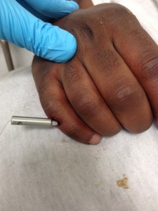
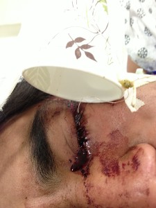
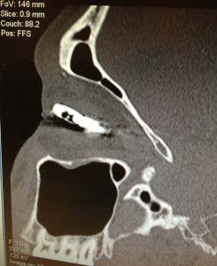
The 3 following cases present to your ER. What should you do?
-A 35 YOM no PMH with a TASER electrode in the eyelid and another to his chest wall, denies chest pain, normal VS
-A 40 YOM PMH polysubstance abuse and schizophrenia who was tasered once to the back is sweating profusely and shrieking incoherently. Triage was unable to take his vital signs due to extreme agitation.
-A 50 YOF PMH CHF w/ AICD with a TASER electrode over her chest wall. She was noted to have fallen 5 feet from a ladder. She has ecchymosis to her face and refuses to move her right arm.
TASERs, the most well known brand of conducted electrical weapons (CEWs), were made popular as a non-lethal weapon alternative to be used by law enforcement to help subdue violent, absconding, or potentially dangerous people. CEWs use compressed nitrogen to propel two electrode darts connected to wires into the skin. They deliver rapid pulses of high voltage and low current that cause involuntary neuromuscular stimulation. This differs from the “stun gun” in which the painful shock requires direct contact between the gun and the skin.
Since become commercially available in 1974, CEWs have been deployed more than 1.5 million times. In parallel with the increasing popularity of these devices, there have been increases in the number of reported deaths in individuals where a CEW was used. Between the years of 2001-2011, there were 466 deaths following CEW use. Not surprisingly, the medical community, politicians, law enforcement, and human rights groups continue to vigorously debate the safety and ethics of these devices.
My focus will be on CEWs as they relate to the Emergency Department including the pathophysiology of injuries, the medical management of patients, and the highly fatal disorder known as Agitation Delirium.
CEW injuries fall into 3 categories: (click to expand explanations)

photo courtesy of Dr. Schechter
1. Primary Barb Injuries
Similar to fishhook injuries, the small barbs on the electrodes typically penetrate the dermal and epidermal layers of the skin, however because of the variation in dermal thickness, for example the dermis in the eyelid is 3mm whereas the dermis in the back is 3 cm, it is possible to injure hypodermal structures. The patient also may have superficial burns between the two barbs. For most tissue injuries, barbs can be removed by applying inward traction while the other hand grips the electrodes and pulls back firmly. Anesthesia is not necessary because of the small caliber of the barbs and its electrocautery effect to the tissue. Ocular injuries can include eyelid trauma, iritis, globe ruptures, macular cysts, and cataracts, and should therefore be emergently referred to an ophthalmologist. Any retained electrodes in the eye should not removed by the ED physician. In addition to the eye, injuries to joints, cranium, or locations where there is a risk of the dart penetrating a body cavity should be removed with care and with the appropriate consultation if warranted. Tetanus prophylaxis should be offered to all patients.
2.Electrical Complications
Cardiac
The standard TASER delivers 5 seconds of low current with short pulses for a total of 2mA with each discharge of the weapon. The threshold to depolarize the cardiac membrane is approximately 100mA. Additionally, current flow takes the path of least resistance, preferring to spread within the superficial tissues and not penetrate to the organs. In 23 different trials in peer-reviewed journals with healthy human volunteers receiving 5 or 10-second CEW applications, ECG or echocardiogram monitoring, showed no ventricular arrhythmias and no changes to troponins, QRS, QT, or QTC. There was also no effect on blood pressure. There were some significant transient changes noted, including increases in heart rate (2.4-15 beats per minute), decreases in PR interval related to the increase in pulse, some nonspecific ST changes, and sinus arrhythmias. The limitation to the studies relying solely on ECGs is that the actual deployment interferes with ECG monitoring. The real-life limitations to relying on these studies is that while 5 seconds is the standard discharge time, in the field, 50% of discharges are “singles”, and 50% of the time discharges are “multiples”. Moreover, these studies were done in individuals without known structural heart disease, outside of a highly stressful environment, and without confounding factors such as drugs or psychosis. Critics of CEWs often cite a famous case of a 16 year-old male with ADD and asthma who immediately fell unresponsive after receiving two 5 second shots to the chest wall. The EMS ECG after 1.5 minutes of CPR showed Vfib and after 4 rounds of defibrillation, the subject was pronounced dead. Pathologists later noted a possible arrhythmogenic right ventricular cardiomyopathy in the patient as well as marijuana in his blood. In animal studies, when the “dart to heart” distance was less than 2 cm and the discharges were given for prolonged periods of time, fatal cardiac arrhythmias were noted in some subjects, for example after two 40 second CEW exposures 3 in 19 pigs in one study had fatal vfib. In 2009, TASER International updated their guidelines to “avoid chest shots” whenever possible as the estimated arrest-related death rate is 1 in 100,000 in “physiologically or metabolically compromised persons.“
In the emergency department, patients with CEW-related cardiac complications will have presented during their pre-hospital care and we will continue ACLS resuscitations. While there is a paucity of evidence for patients with pacemaker or defibrillator devices, most sources recommend interrogating the device prior to making a disposition. In asymptomatic individuals, routine ECG and troponins are not necessary.
Musculoskeletal
CEWs stimulate the A-α motor neurons causing muscle contractions, which can lead to muscle cell damage and the subsequent release of creatinine kinase. In numerous studies CK and myoglobin did increase significantly, but not to a level considered important clinically. Therefore CK is not part of routine laboratory testing.
3. Secondary Injuries
Secondary Trauma
A CEW discharge induces muscle tetany and a loss of muscle tone which may result in two complications 1) falls and 2) tetany-induced bony fractures. Fall injuries often occur from the temporary loss of muscle control while the subject is running, jumping, or climbing. Moreover, the loss of voluntary muscle control may prevent them from maneuvering in such a way to lesson the impact of their fall, such as breaking their fall with an outstretched hand. A complete secondary survey must be conducted to evaluate for trauma.
Agitation Delirium
Agitation delirium is often precipitated by an extreme adrenergic state where an individual on drugs such as cocaine, PCP, or bath salts uses intense physical exertion to resist arrest. If the individual is apprehended, for example, by TASER, handcuffs, or chokehold, this furthers their adrenergic state. Agitation delerium is multifactorial, with alcohol or drugs seen in 70-100% of those with poor outcomes and extreme physical exertion found in 78%. Psychiatric illnesses such as mania or schizophrenia or systemic illnesses were also noted in case reports of patients with agitation delirium. An interesting study in the 1980s was the first of many studies to note that the average cocaine blood levels in patients who died from agitation delirium were lower than levels seen in typical cocaine overdose fatalities suggesting that while cocaine factored into the deaths of these individuals, their cause of death was not from the typical mechanism of a cocaine overdose. Both stimulants and stress trigger excessive dopamine release, which in turn triggers the autonomic hyperactivity seen in patients with agitation delirium, though the exact mechanism is incompletely understood. Many consider agitation delirium to be on the same spectrum as neuroleptic malignant syndrome (NMS). Like NMS, agitation delirium patients may display a sudden onset of paranoia, hyperactive behavior, hyperventilation, and hyperthermia. The mortality rate approaches 84%, with two-thirds dying on scene from respiratory failure or cardiac arrest. Those who survive long enough to reach the emergency department often suffer from complications such as rhabdomyolysis, DIC, and renal failure.
Management should start with rapid sedation. While we may instinctively reach for our trusty neuroleptics (Haldol) and benzodiazepines (Ativan), patients don’t have time to wait the 10-15 minutes for the onset of these medications. IM Ketamine (4-5mg/kg/dose with onset of action in 3-4 minutes) or IV Ketamine (1-2mg/kg/dose with onset of action of 30 seconds) quickly produces sedation and analgesia. Once sedation is achieved, IV, 02, and monitoring should rapidly be established. The autonomic hyperactivity must also be addressed and therapies such as Clonidine and Dantrolene should strongly be considered. Full labs including Creatinine Kinase and blood gases should be obtained.
Summary of ED Management:
- Primary Survey
- If suspect Agitation Delirium, sedate with Ketamine initially, consider Clonidine and Dantrolene. Can use Haldol and Benzos for prolonged sedation. Supportive care. MICU consult. Full labs including CK and blood gases.
- Secondary Survey- Routine labs and ECG not required for asymptomatic healthy patients with normal clinical exam and CEW exposures up to 15 seconds regardless of location of injury.
- Soft tissue injuries should all be offered tetanus prophylaxis. Use firm traction to remove the barbs unless the injury is to the eye or joints. Globe injuries must have emergent ophthalmology consultation. If chest wall injury, consider chest Xray for pneumothorax (especially if patient’s BMI is < 25) or if clinical symptoms present. Joint injuries may require evaluation for joint capsule penetration.
- ICDs or pacemakers should be interrogated, if normal function and no recorded events, can discharge patients; if abnormal, admit to telemetry.
- Evaluate for fractures or other injuries and treat appropriately
- Pregnant over 24 weeks should be referred to OB for cardiotocography
By Dr. Wendy Chan
Works Cited
Bozeman, W.P., Teacher, E., and Winslow, J. “Transcardiac Conducted Electrical Weapon (TASER) Probe Deployments: Incidence and Outcomes.” The Journal of Emergency Medicine 43.6 (2012): 970-75. Web.
Jauchem, J.R., PhD. “Exposures to Conducted Electrical Weapons (Including TASER Devices): How Many and for How Long Are Acceptable?” Journal of Forensic Sciences 60.S1 (2014): S116-129. Print.
Kunz, S. N., N. Grove, and F. Fischer. “Acute Pathophysiological Influences of Conducted Electrical Weapons in Humans: A Review of Current Literature.” Forensic Science International 221.1-4 (2012): n. pag. Web.
Pasquier, M., MD, Pierre-Nicolas C., MD, Laurent V., MD, and Bertrand Y., MD. “Electronic Control Device Exposure: A Review of Morbidity and Mortality.” Annals of Emergency Medicine 58.2 (2011): 178-88. Web.
Megan, R., Close, B., Jeremy Furyk, J., and Aitken, P. “Review Article: Emergency Department Implications of the TASER.” Emergency Medicine Australasia 21.4 (2009): 250-58. Web.
Sztjnkrycer, B., MD. “Cocaine, Excited Delirium and Sudden Unexpected Death.” Journal of Emergency Medicine Service 34.4 (2005): 77-81. Print.
Takeuchi, A., MD, Terence L. Ahern, BA, and Sean O. Henderson, MD. “Excited Delirium.” Western Journal of Emergency Medicine XII.1 (2011): 77-82. Print.
Zipes, D. P. “TASER Electronic Control Devices Can Cause Cardiac Arrest in Humans.” Circulation 129.1 (2014): 101-11. Web.
wendyrollerblades
Latest posts by wendyrollerblades (see all)
- EM-CCM Hematology 7/22/2015:Treating DIC - July 27, 2015
- Critical Care: Recent Trials in Sepsis - May 1, 2015
- TASER Conducted Electrical Weapon Injuries - March 9, 2015
- Racially Biased Pain Control in the ED - December 15, 2014
- Cyra Whaa?!: Cautionary Tales About ER Folks Who Didn’t Use Skilled Translators - September 22, 2014


