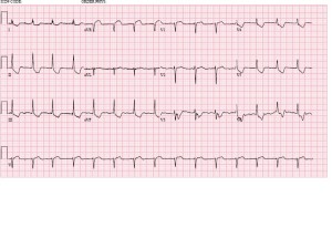
67 yo F with permanent atrial fibrillation. Why is her rhythm regular?
Best answer by May 18th at noon is our winner!
The views expressed on this blog are the author's own and do not reflect the views of their employer. Please read our full disclaimer here. Any references to clinical cases refer to patients treated at a virtual hospital, Janus General Hospital.
The following two tabs change content below.


eabram
Latest posts by eabram (see all)
- Rhythm Nation May 2015 Answer! - June 1, 2015
- Rhythm Nation May 2015 - May 10, 2015
- Rhythm Nation April 2015 – Answer! - April 20, 2015
- Rhythm Nation April 2015 - April 13, 2015
- Rhythm Nation March 2015 – Answer! - March 31, 2015


This can be a n accelerated junctional rhythm due to complete heart block. No atrial pulse is transferred though AV block, so the source of ventricle contraction is the junction, right below the AV node which produces a narrow complex. This rhythm can be seen in dig toxicity.
Otherwise, the rate is 70, normal axis, normal QRS and QT duration, possible STE in AVR and T inversion in I, II, III, AVF, V3-V6, which depending on the clinical picture is concerning for ACS (LM occlusion)
I agree with Reza. Regulization of permanent afib is concerning for complete heart block due to cardioactive steroid toxicity. The ST morphology is probably from the digoxin effect.
An baseline irregular rhythm that converts to regular sinus would be concerning for a complete AV nodal block which then exposes the underlying regular beat inherent to the Ventricles.
New onset AV block could be caused by infarction (predominantly RCA infarcts or less likely circumflex infarcts) or by the presence of AV nodal blocking agents, less likely by lyme disease, or congenitally by lupus in the mother.
However given this pt’s ST morphology ischemia is less likely (no Q waves, no frank ST elevations) and a digitalis effect is more likely. It has the classic ST down-sloping appearance (“Salvador Dali’s moustache” as well as depressions in V4-V5. You can’t really see if the PR intervals are increased on this EKG unless you assume the P wave is buried in the T-waves of each previous beat (which would also imply that the dig has converted the patient our of a-fib). Qt segments appear normal as well, and maybe a hint of a biphasic T-wave in V3 and V4 and V6.