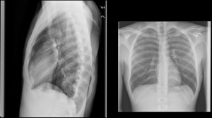
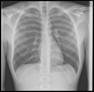
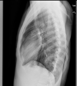
20 year old muscular 6’3″ man presents to the ED with chest pain and states that it feels like “bubble wrap popping” when he bends over and reports some difficulty breathing. No hx of trauma. You get a CXR and see:

 Vitals: BP 110/80, HR 105, RR 22, O2 sat 95% on RA, afebrile. What does he have and how would you manage this?
The views expressed on this blog are the author's own and do not reflect the views of their employer. Please read our full disclaimer here. Any references to clinical cases refer to patients treated at a virtual hospital, Janus General Hospital.
The following two tabs change content below.


sliang
EM-IM Resident at SUNY Downstate/Kings County Hospital
Latest posts by sliang (see all)
- Xray Vision: Chest Pain Answer - June 26, 2015
- Xray Vision: Chest Pain - June 14, 2015

Pneumothorax. Deep sulcus sign on XR.
Management depends on the size. Based on his vitals he appears stable, and the PTX is small or we would see more significant findings on XR.
I would get a CT chest to further characterize the pulm defect and likely observe the patient with a low threshold for placing a chest tube if he decompensates. Admit to surgical ICU for close monitoring and supplemental O2.
Pneumomediastinum. Although pneumomediastinum can occur spontaneously and is for the most part managed conservatively, this patient is symptomatic with abnormal vitals. In addition to “IV, O2, monitor”, I would get a CT chest with contrast to assess for lung pathology and contrast shallow study to assess for esophageal rupture. Depending on the etiology this patient may require ICU level of care.
This is a pneumothorax, complicated with subcutaneous emphysema. The lateral view shows these 2 findings more clearly. The subcutaneous emphysema is seen more apparent on the posterior chest wall.
Correlating this clinically, he is saying he is short of breath with sats of 95% because of this small pneumothorax and the bubble wrap popping is his way of describing crepitus. This is also likely felt on the exam during palpation of the posterior chest wall that has been omitted from the presentation.
A POC ultrasound would be interesting to locate his lung point. I am not sure how subcutaneous emphysema would look like on US, but interesting nonetheless.
Sathya
Peds EM
PGY 4
Sorry forgot that I should mention management.
Chest tube. Admit to ICU. Subcutaneous emphysema will worsen if there is no chest tube in his left lung.
Sathya