Rhythm Nation: Case 4

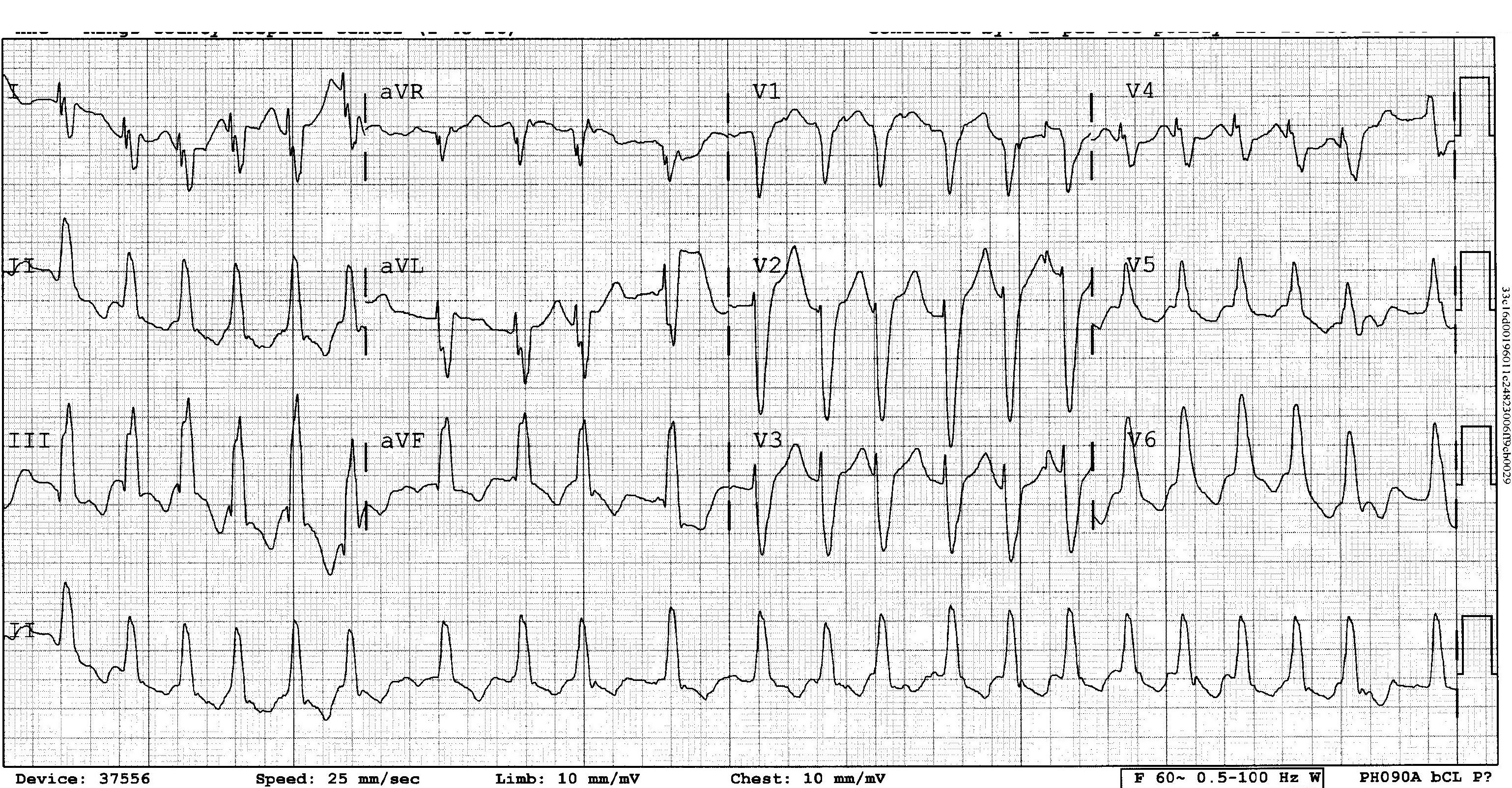
Here’s this months EKG!  As usual… post your comments and questions below…  I will post the answer on Friday!


The views expressed on this blog are the author's own and do not reflect the views of their employer. Please read our full disclaimer here. Any references to clinical cases refer to patients treated at a virtual hospital, Janus General Hospital.
The following two tabs change content below.

nchristopher

Latest posts by nchristopher (see all)

6 comments for “Rhythm Nation: Case 4

  1. Mark Silverberg
    December 11, 2012 at 12:41 am

    If u guys r going to take a stab at this one, remember to be organized. Go through systematically. Large tasks are often easier when broken down into steps.

  2. Nathan
    December 11, 2012 at 1:47 pm

    At first glance this looks like VT, but it is irregular and the complexes in the precordial leads are discordant. This is probably a SVT with abberent conduction.

  3. jfreedman
    December 11, 2012 at 2:13 pm

    This is wide, fast, and irregular rhythm.

    The DDx for that is polymorphic VT, A fib with abberancy, or A fib with WPW.

    p waves: not obviously seen
    Rhythm: Irregularly irregular
    Ventricular Rate: 132bpm
    Axis : RAD, LPFB?
    QRS morphology: LBBB
    Intervals:QRS is not exceptionally wide; QT is looks kinda long (but its fast)
    ST/T: c/w LBBB

    For this to be polymorphic VT, we’d see QRS of varying amplitudes, which we don’t. IT’s not fast enough that WPW is on my radar. I think this is a fib with RVR, LBBB.

    So this is

  4. melton
    December 12, 2012 at 5:19 pm

    I agree with Nathan and Joey, this is a wide complex tachycardia of supraventricular origin. There are a host of reasons why this is not wide complex VT.

    Some things that would make this more likely WCT VT include:

    fusion beats
    av dissociation
    leads V1-V6 pointed in the same direction (concordant)
    LAD
    QRS duration in RBBB >140 msec or LBBB >160 msec
    capture beats
    evidence of old MI ie. q waves

  5. Carl
    December 14, 2012 at 1:16 pm

    I agree with everyone with one exception.
    I think I see P waves which march out at a rate of ~300. This is a wide complex, irregularly irregular. However most of this strip is in a regular rhythm, with occasional areas of abnormal beats.

    In addition to the prior differential this could be a pacemaker tachycardia, and electrolyte arrythmia.

    I think I see a lot of P’s and the PR ratio looks almost fixed, but very short, perhaps with an element of wpw here. The atrial rate of 300 makes me almost certain this is aflutter.

    I think this is an atrial flutter rhythm with a variable block, and LBBB conduction delay. I’m not sure about an accessory pathway, but the PR sure is short.

  6. Nordstrom
    December 15, 2012 at 10:20 pm

    Here’s my guess.

    Rate: ~150
    Rhythm: see below
    Axis: Right

    P waves: yes, but abnormal and difficult to characterize with the tachycardia
    PR: Variable
    QRS: wide (but not obviously) 0.12 to 0.14 sec, LBBB like pattern
    QT: nml, 0.36 to 0.40 sec

    Overall my guess is a ventricular tachycardia. My main reason is multiple spots where I think I see P waves with AV dissociation. The QRS has a LBBB like pattern, but I don’t think its a supraventricularly driven tachycardia with a LBBB block. I agree with Jay, the other signs of Ventricular Tachycardia that he mentioned are not present.

Comments are closed.