Rhythm Nation: Not Just the EKG

photo (5)Here’s this weeks EKG case!  Thanks to Niki for submitting the case!

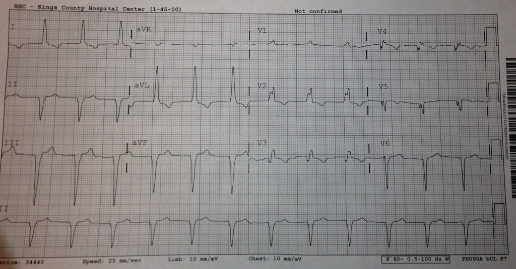
This week we’ll try to not just interpret the EKG but use it in addition to other clinical findings to bring us to our diagnosis.

59yo F presents with a chief complaint of chest pain and shortness of breath.  Her triage vitals are 157/102, 84, 20, 97.4, 94% sat on RA.

Her only hx that she can recall is HTN and arthritis.  She is visiting from the West Indies.  There are no prior EKGs available.

Please reply below with your interpretation of the EKG.  Additionally, you can ask for further history, exam findings, labs, imaging, ultrasound… etc…  How would you work up this patient?  What studies are essential? What is your differential?


The views expressed on this blog are the author's own and do not reflect the views of their employer. Please read our full disclaimer here. Any references to clinical cases refer to patients treated at a virtual hospital, Janus General Hospital.
The following two tabs change content below.

nchristopher

Latest posts by nchristopher (see all)

2 comments for “Rhythm Nation: Not Just the EKG

  1. andygrock
    April 25, 2013 at 7:03 am

    Rate: 70ish

    Rhythm: Regular

    Axis: extreme L axis deviation!!!

    Intervals: unk PR (see below), nml QT, wide QRS

    P waves: no visible p waves….possibly visible p wave in III/AVF. Perhaps buried in qrs? perhaps no p wave?

    QRS: wide complex, RSR’ in V2 c/w RBBB. Large, upright qrs in I/AVL c/w LBBB. RS in II/III/AVF c/w LAFB (but no qr in I/AVL that you would expect with LAFB)

    T wave: inverted in I/AVL/V2/V3/V4/V5

    No electrical alternans

    ST segments: small ST depressions in I/AVL (<1mm). ST elevations (look like j point elevations) in II/III/AVF.

    Put it all together: Trifasicular block…again?
    MI with AV dissociation.
    Per Dr. Martindale inverted T waves in precordial leads can indicate PE.

    This is a tough EKG.
    Her story sounds PEish (who knows if she's actually tachypnic…everyone is 20.)
    I want: IV/O2/Monitor, old ekg, VBG, bedside echo by Dr. Schecter, old ekg, CXR, CBC/cmp/trop/d dimer (if you say PE number 1 dx then wells 3, moderate risk, jump to CTA). Also, let's do a physical exam including but not limited to heart/lungs/lower extremities.

    Differential: PE,PE, PE (recent travel – visiting from west indies/low sat/cp/sob)
    Pulmonary artery hypertension (ILD/genetic/OSA etc)
    MI leading to complete heart block/trifasicular block
    CHF exacerbation with strange ekg at baseline.
    Dig toxicity (maybe she's drinking the foxglove plant – is she seeing everything yellow/with halos?)
    hyperkalemia

    Well, 7AM, night shift over. I guess I should sign off now.

  2. nchristopher
    April 26, 2013 at 7:54 am

    Thanks for the thorough reply.

    The positive findings of the physical exam are as follows:
    CV: nml S1/S2; holosystolic murmur heard at the L sternal border 3/6;
    Chest: Coarse but equal breath sounds bilaterally
    Abd: Soft; NT/ND; nml BS
    Extr: Edema B/L slightly more on LLE

    Again there is no prior EKG available!

    Dr. Schecter’s shift doesn’t start for a few hours… so I will post the echo soon!

Comments are closed.