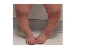
22 months old previously healthy girl was brought to the ED by her parents with chief complaint of limping on right side while walking and running for 1 day. They noticed this after she fell at home while running. Parents denied any fever, runny nose, cough, rash and joint swelling. On physical exam, she was well appearing. Slight limping on right side appreciated with walking and the following leg abnormality was noted:
Picture:
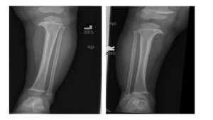
Her x-ray of pelvis and femur was within normal limits. X-ray of tibia and fibula showed the following abnormality:
What is your diagnosis?
Answer: Blount disease (also called Idiopathic tibia vara)
It is a developmental deformity resulting from abnormal endochondral ossification of the medial aspect of the proximal tibial physis leading to varus angulation and medial rotation of the tibia.
Etiology
Largely unknown, though likely multifactorial (genetic, environmental and mechanical factors interplay).
Types
Three types: infantile (1-3 yr), juvenile (4-10 yr), and adolescent (11 yr or older)
The juvenile and adolescent forms are commonly combined as late-onset tibia vara.
Infantile form:
-Predominant in black girls
-Usually bilateral (80 percent of cases)
-Greatest potential for progression
The juvenile and adolescent forms (late–onset):
– Predominant in black boys
– Approximately 50% bilateral involvement
– Slowly progressive
Risk Factors
-Female sex
-African-American race
-Obesity
-Early walking
-Positive family history
Clinical presentation
-Bowing of the leg below the knee (varus deformity)
-Leg-length discrepancy
-Knee pain
-Limping
-Intoeing
-Asymmetric angular alignment of the lower extremities
-Focal angulation at the proximal tibia
-Lateral thrust (a brief lateral knee-joint protrusion) during the stance phase of ambulation
Diagnosis
An anteroposterior standing radiograph of both lower extremities with patellas facing forward and a lateral radiograph of the involved extremity should be obtained. Weight-bearing radiographs are preferred and allow maximal presentation of the clinical deformity.
The characteristic radiographic features of Blount disease include medial beaking and downward slope of the proximal tibial metaphysis.
Management
Depends upon the age of the child and the severity of the deformity.
Patients with infantile Blount disease may be treated with braces to unload medial compressive forces. Brace therapy is successful in 50 to 80 percent of patients. If the deformity does not resolve with bracing, surgical correction is needed.
In late-onset tibia vara, bracing is usually ineffective, surgical correction is the mainstay of treatment.
References
Kliegman RM, Stanton BF, et al. Nelson Textbook of Pediatrics. 19th edition. 2011; 2344-2351
Zionts LE, Shean CJ. Brace treatment of early infantile tibia vara. J Pediatr Orthop 1998; 18:102
Bradway JK, Klassen RA, Peterson HA. Blount disease: a review of the English literature. J Pediatr Orthop 1987; 7:472
marmeg
Latest posts by marmeg (see all)
- Let’s pretend that an imaginary patient comes into your pediatric ED… - June 8, 2015
- Foreign Body Ingestions in Children, by Abi Iyanone - March 15, 2015
- Just another case of vomiting? By Dr Subramaniam - February 3, 2015
- Limping, continued… By Nishit Patel MD - December 29, 2014
- Approach to the Limping Child by Dr. Virteeka Sinha - November 12, 2014


